Artistic Anatomy · Part 2 of 5 · May 30, 2021
Artistic Anatomy #2 Torso, Front Layers
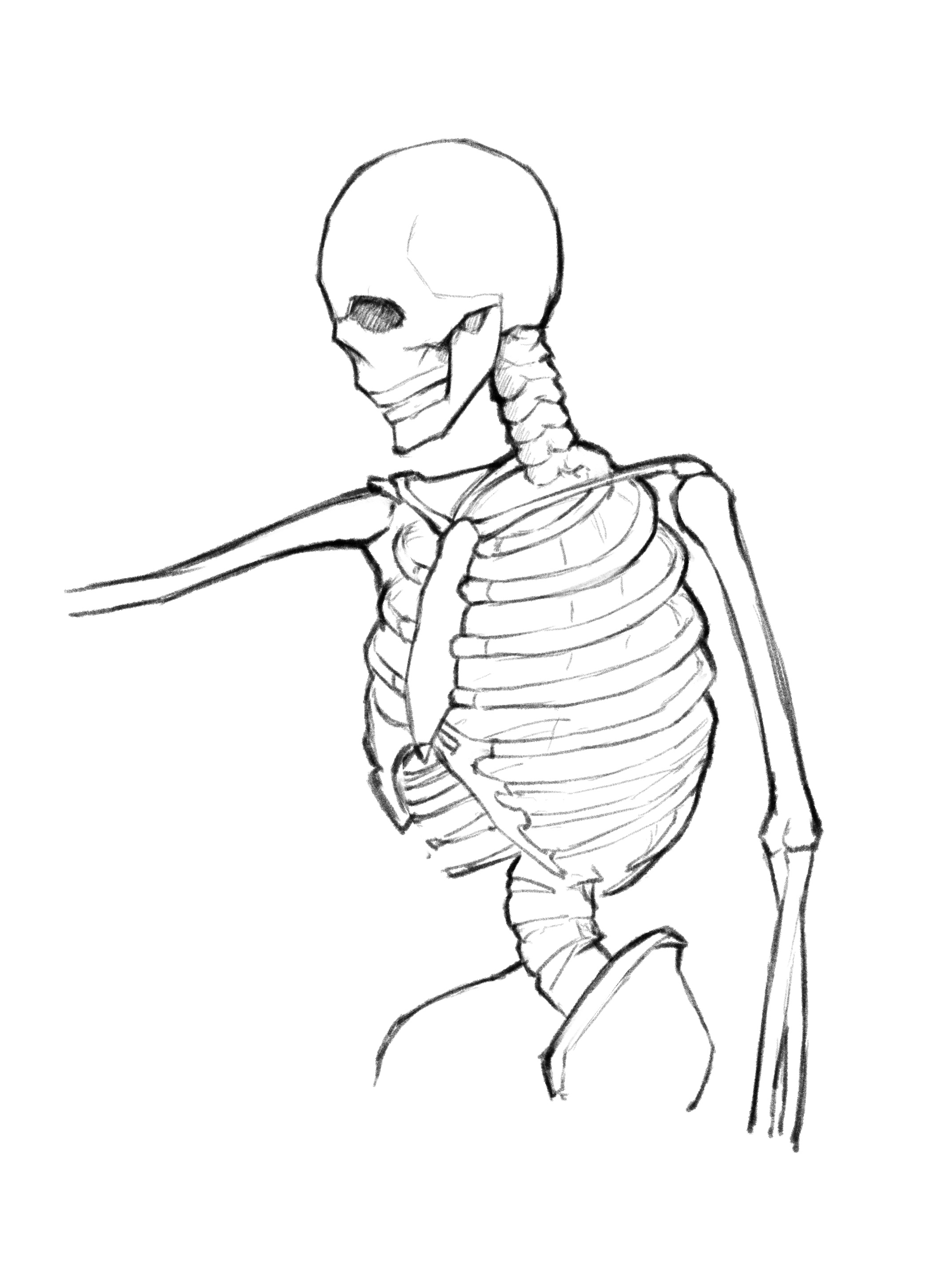
This note covers the layering relationships of the main muscles on the front of the torso.
The previous note went through the torso muscles one by one, recording each origin and insertion. But when drawing, what you actually need is not a list of muscles — it is the order: which one sits underneath, which one covers it, and who overlaps whom at the boundary. Get the layering backwards and the form is wrong immediately, even if every individual muscle is correct.
Starting from an empty skeleton. The skeletal structure drawn here is not strictly accurate and serves only as a positional reference. Only a few bony landmarks are needed: the clavicle, the sternum, the lower edge of the ribcage, the ASIS and the humerus. Everything hangs off those.
The layering order
From deep to shallow, the front is four layers:
- On the bone — serratus anterior and external oblique. They grow directly on the ribs and form the floor for everything else.
- The connector — rectus abdominis, filling the gap between the lower ribcage and the pubis.
- The cover — pectoralis major and latissimus dorsi. Pec major covers the upper half of serratus from above; lats wrap around from behind and cover its lower half.
- The outer layer — deltoid and trapezius. They cross the shoulder and hide everything converging onto the humerus.
Build in that order and the boundaries come out right. Each layer follows.
Serratus anterior
- The serratus anterior lies on the ribs and can be approximated as wrapping around them from back to front, unit by unit.
- The serratus anterior should not extend too far. From the front view it sits on the sides of the body; no serrations should appear near the centre line.
- Generally only lean or muscular people show clearly visible serratus lines on the surface.
There is a common mistake here: drawing all the serrations. Serratus anterior originates on the upper 8–9 ribs, but the upper ones are covered by pec major and lats, so usually only the bottom 3–4 are visible on the surface. Drawing more makes the ribcage look too wide.
Note also that the serrations point diagonally back and up, because they all converge on the medial border of the scapula. Drawn horizontally they read as ribs instead.
External oblique
It runs diagonally, and it interlocks with the serratus anterior.
That interlock is the easiest thing to muddle on the front. The serratus teeth point forward and down; the external oblique teeth point forward and up, and the two mesh like a zipper along the side of the body. Remember the direction of the mesh and that whole area stops being a blur.
The external oblique originates on the lower eight ribs and inserts on the iliac crest. Above the ASIS it forms a distinct diagonal mass — the inverted V many people have at the side of the waist is where it meets the rectus abdominis.
Rectus abdominis
The navel sits where the third and fourth segments meet. Pay attention to the tilt, angle and curvature of the muscle.
A few more things worth watching:
- The rectus abdominis is not a flat slab. It is higher in the middle and lower at the sides, curved across its width, and the whole run from sternum to pubis bows forward. Draw it flat and it has no volume.
- The four segments are not equal in width or height: they narrow going down, and the lowest one is the longest. Dividing the area into four equal boxes is the most common error.
- The tendinous intersections separating them are not straight lines; they arch slightly upward. The left and right sides do not necessarily line up either.
- The linea alba runs from below the sternum to the pubis, fading out below the navel. It does not interlock with the external oblique — that boundary is clean.
Pectoralis major
Pec major divides into three parts. The first is triangular, with one side along half the clavicle and its corresponding point on the humerus. The second has one side on the sternum, with its point also on the humerus, slightly above the previous one. The third is as shown in the figure and is difficult to describe in words.
The three bundles' insertions on the humerus overlap, and in reverse order: the clavicular part lands lowest, the sternocostal part in the middle, the abdominal part highest. This is why the front edge of the armpit twists when the arm is raised — the fibres cross over each other at the insertion. It is barely visible from the front, being under the deltoid, but it determines the shape of the anterior axillary wall.
The most common problem when drawing pec major is making the lower edge horizontal. In fact it runs diagonally from high on the outside to low on the inside, and its outer end is clipped by the deltoid.
Latissimus dorsi
The lats extend from the back, so they are discussed in more detail in the back muscles note. For now it is enough to know that the lats wrap around the serratus anterior, and the part visible from the front lies below pec major.
In one sentence: the lats define the rear edge of the armpit. With the arm down they are almost invisible; with the arm raised, a clear diagonal edge wraps around from the side into view. On someone with developed lats, that edge pushes the whole outer silhouette of the torso out — the V-taper is the lats and the deltoid together.
Trapezius
It originates at the mastoid process and extends to the outer end of the clavicle, covering the neck like a cape.
From the front, only the topmost part of the trapezius shows: descending from the side of the neck and inserting on the outer third of the clavicle. The triangular hollow it forms with the sternocleidomastoid — the supraclavicular fossa — is one of the most important structures at the base of the neck, and should not be filled in.
Sternocleidomastoid
Strictly it belongs to the neck, but from the front it is the most conspicuous shape along the top edge of the torso, so it cannot be skipped.
It leaves the mastoid process behind the ear and splits into two heads going down: the sternal head inserting on the manubrium, the clavicular head on the inner clavicle. The two separate at the bottom, leaving a small triangular hollow between them. Turning the head pulls the sternal head into a taut diagonal cord, which is the single most useful structural line when drawing a turned head.
Deltoid
The deltoid has three heads, essentially wrapping around the acromion from front to back, with all insertions on the lateral side of the humerus. Under the deltoid's cover, the point where pec major converges on the humerus is no longer visible from the outside.
Between the deltoid and pec major runs the delto-pectoral groove — a furrow descending diagonally from the middle-outer clavicle to near the deltoid insertion. It is present on every body type and is the most reliable structural line on the shoulder. Many people draw the deltoid and pec major as one continuous mass, and the form falls apart.
Summary
Drawn this way, the front comes down to three things:
- Serratus and external oblique mesh along the side, with their teeth pointing in opposite directions.
- Pec major covers the upper serratus, the lats cover the lower, leaving a band of serrations visible between them.
- The deltoid covers everything converging on the humerus, leaving only the delto-pectoral groove.
The next note moves to the back.