Artistic Anatomy · Part 3 of 5 · June 13, 2021
Artistic Anatomy #3 Torso, Back Layers
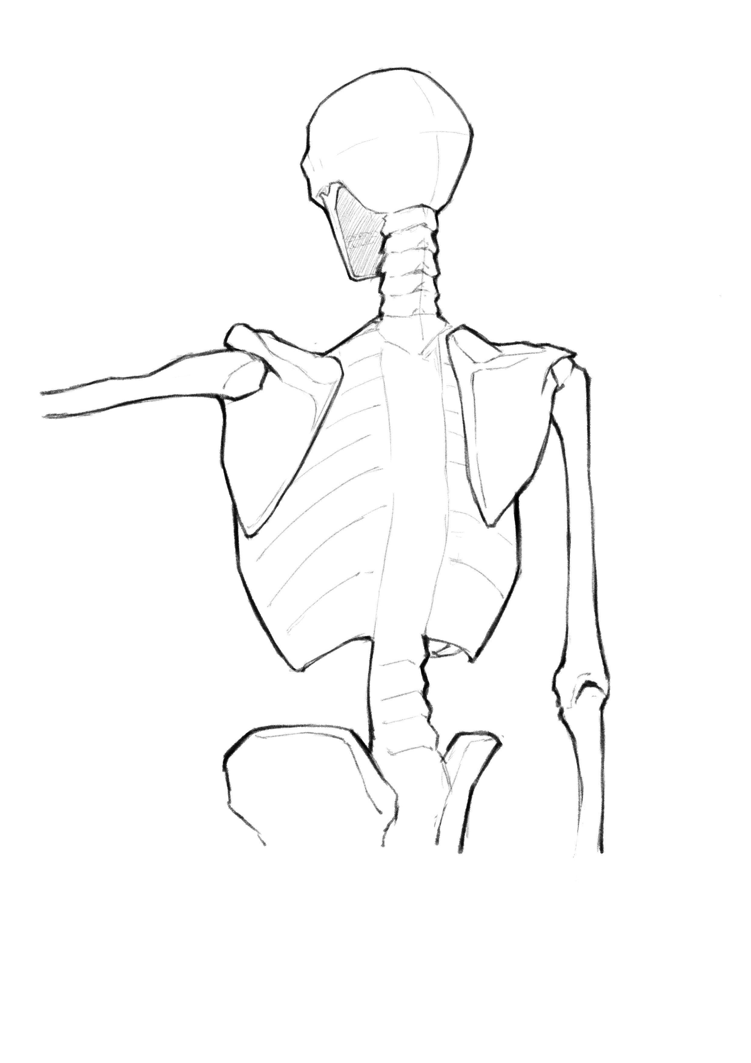
This note covers the main back muscle structures and their layering relationships. The reference is still Anatomy for Sculptors by Uldis Zarins et al.
The skeleton used is shown below. Since the specific distribution and count of ribs and vertebrae are not very helpful for memorizing the main muscles — and are rarely useful except in very hardcore drawing scenarios — the ribcage and spine are represented with simplified shapes in this note.
The back is harder to draw than the front, for two reasons. First, the scapula moves: unlike the ribcage it is not a fixed landmark. Second, there are more layers — trapezius and latissimus cover nearly all the deep muscles, but not completely, and the parts left exposed are exactly what gives the back its shape.
The layering order
From deep to shallow:
- On the bone — serratus anterior, external oblique (the part coming round from the side), erector spinae.
- The scapular layer — infraspinatus, teres minor, teres major, rhomboids. These tie the scapula to the humerus.
- The cover — latissimus dorsi, wrapping from the waist up into the armpit.
- The outer layer — trapezius and posterior deltoid.
Layer 2 is the crux: the rhomboids are covered completely by the trapezius, while infraspinatus and the teres muscles are only partly covered — and the part left showing is that famous bulge below and lateral to the scapula.
Serratus anterior
As shown in the figure, viewed from the back it is largely hidden below the scapula; the undulations can be observed from the sides. On lean or muscular people the distinctly serrated appearance is visible on the surface from the side.
Worth noting: serratus anterior inserts on the medial border of the scapula. It is the muscle that holds the scapula down against the ribcage. When it fails the scapula lifts away (winged scapula), so letting the medial border float slightly off the back is an effective way to draw a relaxed or frail figure.
External oblique
The external obliques originate from the back of the ribs, so part of them can be seen even from behind.
From the back they appear in the small triangular region between the lower edge of the latissimus and the iliac crest. That region, together with the two vertical ridges the erector spinae raises at the waist, forms the shape of the lumbar transition — the dimples of Venus sit around here.
Erector spinae
The erector spinae runs vertically along both sides of the spine, from the sacrum up into the neck. It is the only major longitudinal muscle group on the back; everything else runs diagonally or across.
For drawing, what matters is not its subdivisions (iliocostalis, longissimus, spinalis) but its overall effect: the spine is not a ridge, it is a groove. The erector spinae forms the ridges on either side of it. The groove is deepest at the waist and shallows going up between the scapulae, where the trapezius covers it.
Drawing the spine as a raised line is the most common beginner mistake. Unless the figure is extremely lean or bending forward, the centre of the back is always a furrow.
Rhomboids
The rhomboids connect the spine to the medial border of the scapula, pulling the two scapulae towards each other.
They are completely covered by the trapezius and never visible on the surface. But they determine where the medial border of the scapula sits: shrugging or squeezing the shoulder blades together is the rhomboids working. You do not draw them, but you need to know that the line of the medial border shifts with the pose rather than staying put.
Infraspinatus
The infraspinatus sits below the spine of the scapula, which is where its name comes from.
Origin: the back of the scapula below the scapular spine Insertion: the middle of the greater tubercle of the humerus
Its upper part is covered by the trapezius and its outer part by the posterior deltoid, so what shows is a diamond-shaped region in the middle and lower area. On a muscular figure, that region together with teres major and minor forms a clear mass below and lateral to the scapula.
Teres major and teres minor
Both the teres major and teres minor connect the scapula and the humerus; their layering relationship is shown in the figure below. Note the positions where each connects to the humerus. In practice, the teres minor is difficult to observe on the body surface due to coverage by the infraspinatus and deltoid, but it can still be clearly seen from certain angles.
Concretely:
- Teres minor originates around the midpoint of the lateral border of the scapula and inserts on the lower part of the greater tubercle of the humerus. It lands on the same side as the infraspinatus.
- Teres major originates at the inferior angle of the scapula and has to wrap round to the front of the humerus, inserting near the bicipital groove.
So teres major and [infraspinatus + teres minor] sandwich the humerus from opposite sides. That sandwich is the key to the rear edge of the armpit: with the arm raised, teres major and latissimus together form the thick posterior wall of the axilla.
The three muscles together make one mass below and lateral to the scapula. When drawing the back it helps to block that mass in as a single volume first, and only then divide it into three.
Latissimus dorsi
The latissimus is the largest single muscle on the back and the main determinant of its outer silhouette.
Origin: lower thoracic and lumbar vertebrae, sacrum and iliac crest, through a large aponeurosis Insertion: the front of the humerus, at the bicipital groove
Key points:
- It wraps from below upward and from behind forward. The lower end is very wide, spanning nearly the whole waist; it narrows going up, passes round the side of the body, and finally twists into a narrow band disappearing into the armpit. That twist is the hardest part of its form to draw.
- It covers the lower half of serratus anterior, the upper part of the external oblique, and the region around the inferior angle of the scapula.
- Between its upper edge and the lower edge of the trapezius a triangular area is left exposed, showing the inferior angle of the scapula and teres major. On lean figures this triangle is obvious, and it is a good check on whether the back is constructed correctly.
- Its outer edge forms a diagonal running down from the armpit along the side of the body. That edge plus the deltoid is the outline of the V-taper.
Trapezius
The trapezius is the cape laid over everything else, running from the base of the skull down to the lower thoracic spine.
Origin: occipital bone, nuchal ligament, cervical and thoracic spinous processes — note C7, which is a visible bony landmark on the surface Insertion: outer third of the clavicle, acromion, spine of the scapula
Its three parts run in completely different directions:
- The upper part descends diagonally outward from the neck to the clavicle. It defines the slope between neck and shoulder, and the angle of that slope is the most direct factor in whether a figure reads as powerful or slight.
- The middle part runs roughly horizontally to the acromion and scapular spine.
- The lower part ascends diagonally outward from the thoracic spine to the inner end of the scapular spine, forming a downward point at the centre of the back.
Watch the length: that lower point is not at the waist but at the middle-to-lower thoracic spine, roughly a little below the inferior angle of the scapula. Drawn too long, the figure loses its waist.
Posterior deltoid
The posterior deltoid originates on the lower edge of the scapular spine and inserts on the lateral humerus. From the back there is a clear boundary between it and the infraspinatus/teres minor.
It is the transition between back and arm. The easy mistake is merging it with the infraspinatus mass — the groove between them has to stay, or the shoulder and the back fuse into one shape.
Summary
A way to remember the back: one groove, one mass, one cape, one wrap.
- One groove: the erector spinae either side of the spine — the centre line is concave.
- One mass: infraspinatus plus the teres muscles, below and lateral to the scapula.
- One cape: the trapezius, from the skull to the lower thoracic spine.
- One wrap: the latissimus, from the waist into the armpit.
Get the relationship between those four right and the back stands up. Everything else is detail laid on top of them.
The next note moves on to the upper limb.