This note mainly covers muscle overlap and basic skeletal structure of the torso. The English version will be updated later. Reference books used: Oxford Artistic Human Anatomy (by Eliot Goldfinger, translated by Li Huijuan, Shanghai People's Fine Arts Publishing House, 1st edition August 2018) and Artistic Human Anatomy (Electronic Industry Press).
As this is artistic anatomy only, it does not cover deep muscles that do not affect surface form. Muscle overlap is described qualitatively; please point out any errors.
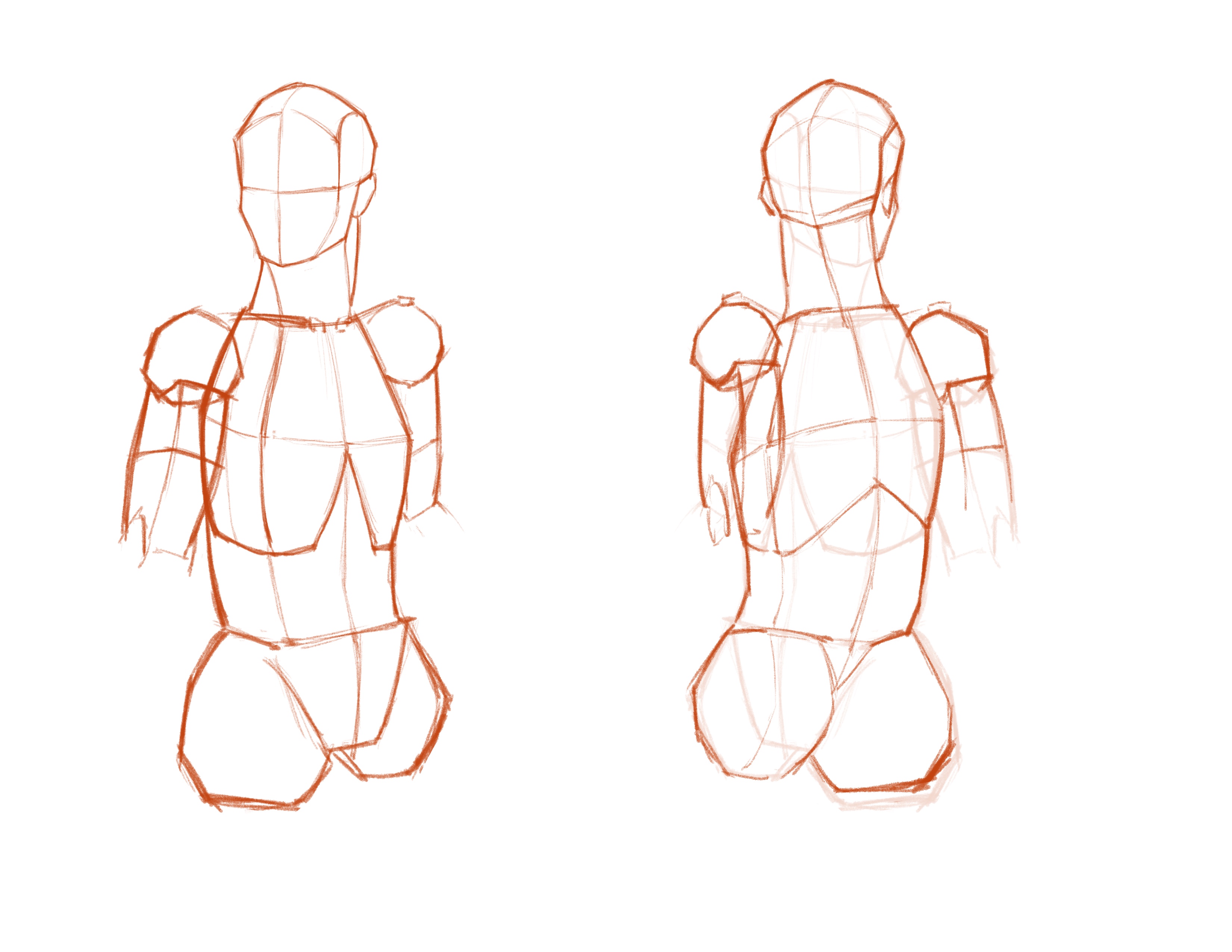
Small Angle · Upper Body Summary
As in previous notes, we still summarize the head, thoracic cavity, pelvic cavity, clavicle, hips, scapula, and shoulder ball. We use the following poses to observe upper body muscle overlap. Start by sketching roughly as shown below.
Sternocleidomastoid
Starting with the head and neck: although there are many complex muscles in this region, few directly and visibly affect surface form. Among them, the Sternocleidomastoid (Stm) is very important. It has two parts: the clavicular head and the sternal head.
Sternocleidomastoid – Sternal Head (SC)
Origin: Mastoid process (can be understood as behind the ear)
Insertion: Sternum
Diagram:
Sternocleidomastoid – Clavicular Head (SS)
Insertion: Clavicle
From the surface, the sternal head is more visible, especially when turning the head.
Trapezius
The Trapezius (Tr) is another important muscle in the head and neck region. It is a large muscle that is clearly visible on the surface.
Origin: Base of skull, cervical vertebrae (note the position of the seventh cervical vertebra C7)
Insertion: Outer third of clavicle, superior border of spine of scapula, tubercle of spine of scapula (roughly at the midpoint of the spine of scapula), base of thoracic cage
Rhomboids
The Rhomboids (Rh) can be divided into rhomboid major and rhomboid minor, but in practice they are barely separable, so treating them as one muscle is fine. The rhomboids are almost never visible on the surface—they are deep muscles. They are not visible from the front.
Origin: Lower part of nuchal ligament (roughly from the end of the cervical spine—i.e., near C7) down to the fifth thoracic vertebra
Insertion: Medial border of scapula
Diagram: The rhomboids lie beneath the trapezius. For clarity, the trapezius is shown with transparency here.
Infraspinatus
The Infraspinatus (In) is a muscle of the back. It lies below the spine of the scapula, hence its name.
Origin: Below the spine of scapula
Insertion: Middle facet of greater tubercle of humerus (the greater tubercle on the upper arm bone)
Diagram: The infraspinatus also lies beneath the trapezius. For clarity, the trapezius is shown with transparency here.
Teres Major and Teres Minor
Teres Major (TMa) and Teres Minor (TMi) are both muscles between the scapula and humerus. The infraspinatus and teres major sandwich the teres minor to form the infraspinatus region of the back. Note that teres minor and infraspinatus cover the same side of the greater tubercle of the humerus, while teres major wraps around the humerus and inserts near the biceps. So teres major and teres minor/infraspinatus grip the humerus between them.
Teres Minor (TMi)
Origin: Roughly the midpoint of the lateral border of the scapula
Insertion: Inferior facet of greater tubercle of humerus
Only a small part of teres minor is visible on the surface.
Teres Major (TMa)
Origin: Inferior angle of scapula
Insertion: Humerus, roughly at the biceps position
Pectoralis Major
The Pectoralis Major (PMa) is a very visible surface muscle between the clavicle and ribs. Although it is one muscle, its structure can be described as three parts: clavicular, sternocostal, and costal-abdominal.
Clavicular Part
Origin: Inferior border of clavicle (medial half of clavicle)
Insertion: Anterior aspect of humerus
Sternocostal Part
Origin: Sternum
Insertion: Anterior aspect of humerus, inserting higher than the clavicular part
Costal-Abdominal Part
Origin: Ribs
Insertion: Anterior aspect of humerus, inserting lower than the sternocostal part
Deltoid
The Deltoid is the most prominent muscle of the shoulder and is clearly visible in motion. Its three heads roughly follow the axis of clavicle–acromion–spine of scapula and insert on the lateral humerus. The anterior head runs from the outer third of the clavicle to the lateral humerus; the middle head from the acromion to the lateral humerus; the posterior head from the lateral spine of scapula, also to the lateral humerus.
Origin: Outer third of clavicle, acromion, inferior border of spine of scapula
Insertion: Midpoint of lateral humerus
Rectus Abdominis
The Rectus Abdominis (RA) is the visible abdominal muscle—the "six-pack" or "eight-pack" refers to the 4 segments on each side of the rectus abdominis. The lowest segment is larger; some people can further divide it into two, but this is rarely visible on the surface. The segments on both sides meet at the linea alba in the middle, separated from the external oblique. The linea alba runs from below the sternum to the pubis and gradually fades below the navel.
Serratus Anterior
The Serratus Anterior lies on the sides of the body. It is named for its saw-tooth appearance. It is more visible in people with lower body fat. It runs along the ribs, originating from the upper 8–9 ribs, but due to muscle overlap, usually only the lowest 3–4 segments are visible on the surface.
Origin: Along the upper 8–9 ribs
Insertion: Medial border of scapula
External Oblique
The External Oblique (EO) adjoins the serratus anterior with interlocking fibers; it borders the rectus abdominis but does not interlock with it.
Origin: Lower 8 ribs
Insertion: Anatomically the external oblique inserts at the linea alba, but for drawing, since the rectus abdominis covers it, we can roughly understand it as ending at the lateral edge of the rectus abdominis.
Latissimus Dorsi
The Latissimus Dorsi (L) is the most prominent muscle of the back. It lies below the trapezius, covers the dorsal part of the external oblique, and reveals the overlap of infraspinatus, teres minor, and teres major. The latissimus wraps from the back to the armpit, so in people with well-developed lats, part of the latissimus contour can be seen from the front. This is especially obvious when raising the arm.