This note mainly records a comprehensive description of muscle interweaving in the upper limb. Due to the extensive content on the upper limb, this note will be continuously updated, and the accompanying figures may be redrawn.
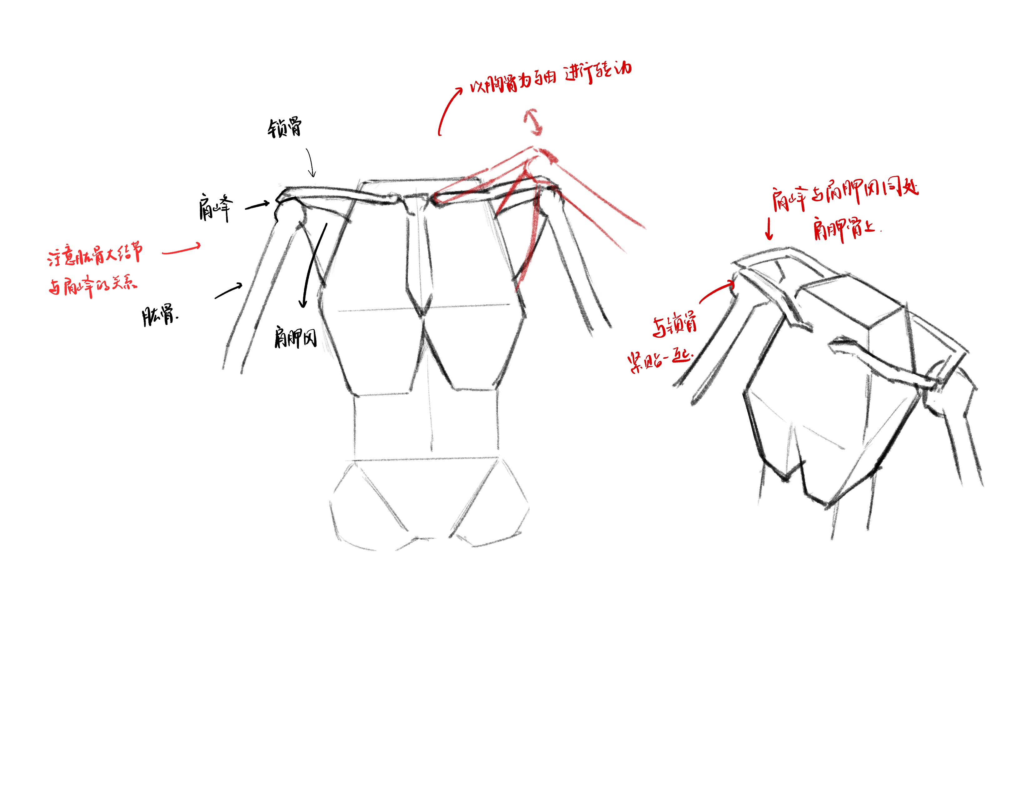
Clavicle-Acromion-Scapular Spine Axis
To understand the movement patterns of the upper limb, we must first return to the deltoid. In common perception, the arm extends from the shoulder down to the palm. In fact, arm movement is inseparable from the shoulder.
After understanding the skeletal relationships of the shoulder, we also need to understand the bone structure of the arm.
Humerus-[Ulna-Radius]
We already know that the bone of the upper arm is the humerus (Humerus). However, the forearm has two bones: one called the ulna (Ulna) and one called the radius (Radius).
Both the ulna and radius connect to the lower end of the humerus, at the elbow joint. The radius is the bone on the same side as the thumb; the other is the ulna.
Schematic diagram:
These two bones are crucial for understanding forearm structure. As shown in the figure above, the radius and ulna cross when the arm rotates. Regardless of how they cross, the radius is always the bone on the thumb side, and the ulna is always the bone on the pinky side. In fact, the bony prominence of the ulna on the wrist side—what people often call the wrist bone—is the clearly palpable tubercle on the hand.
Particularly note that the shapes of these two bones are actually centrally symmetric. When hanging naturally, the radius is smaller at the top and larger at the bottom, while the ulna is smaller at the bottom and larger at the top. The rectangular interface at the wrist is mainly due to the radius being larger on the wrist side. The rotation position with the palm facing forward—where the radius and ulna do not cross—is called supination. The position with the back of the hand forward and the radius and ulna crossed is called pronation. In the figure below, the leftmost is supination and the rightmost is pronation. The middle shows the position with the thumb web facing forward, where the radius and ulna begin to cross but do not form an X shape; this position can also be called semi-pronation.
There's a trick to remember their English names. When holding a bowl of soup with one hand, the arm is in supination. If you then adjust the arm to pronation, the soup will spill, creating a pro-blem—hence pronation is called pro-nation.
It should also be emphasized that during forearm rotation, only the radius and ulna rotate; the position of the humerus does not change.
Again, the positions and shapes of these three bones are very important for understanding the arm! Once fully understood, we can proceed to learning the arm muscles. We will also understand the arm muscles based on the three positions described above.
Deltoid
Regarding the deltoid of the shoulder, we will not elaborate further. The key point is that its three bundles converge at the lateral 1/2 point of the humerus. This is a very important point, as three muscle bundles are related to it. The three bundles attach to the clavicle, acromion, and scapular spine. The greater tubercle of the humerus rests on the glenoid of the scapula and can be roughly understood as being directly below the acromion.
The state of the deltoid also differs in the three natural arm positions. Because the deltoid converges at the lateral 1/2 point of the humerus, forearm rotation causes part of the muscle to rotate. Therefore, even though the deltoid always converges at this point, the middle bundle of the deltoid will be more visible in the pronated state. This is a small detail; in fact, it may not even need to be depicted when summarizing for drawing.
Biceps Brachii
The biceps brachii (Biceps Brachii) is perhaps one of the most easily observable muscles on the arm and the most well-known. When flexing the arm to show muscle, the bulge of the biceps is always taken as a sign of strength.
As its name suggests, the biceps has two origins: one at the supraglenoid tubercle of the scapula and one at the coracoid process of the scapula. It also has two insertions. The lower end of the biceps extends like two bands: one attaches to the radial tuberosity at the upper end of the radius (the prominence at the thinner end of the radius), and one attaches to the middle fascia on the medial side of the forearm (in supination) and wraps around the muscles along the way. Therefore, the naming of the biceps is interesting because it actually has no connection with the humerus. Due to coverage by the deltoid and pectoralis major, the upper part cannot be seen, so when drawing it can be depicted as a mass on the humerus.
Note that in the three rotation states of the arm, although the humerus does not displace, the shape and position of the biceps still change due to the movement of the forearm muscles.
The biceps and deltoid mutually obscure each other. On one hand, the deltoid covers the two heads of the biceps, making it appear from the outside as a simple bulge. On the other hand, all three bundles of the deltoid converge at the lateral 1/2 point of the humerus, passing through from below the biceps.
Brachialis-Coracobrachialis
Both the brachialis and coracobrachialis are closely related to the 1/2 point of the humerus.
The brachialis originates from the front of the humerus at the 1/2 point (in supination) and inserts at the ulnar tuberosity at the elbow joint (note that one band of the biceps attaches to the radial tuberosity at the elbow). Therefore, the brachialis is largely covered by the biceps, pressed beneath it. However, because the brachialis is slightly wider than the biceps, it can still be seen.
The coracobrachialis is different. It originates from the medial side of the humerus at the 1/2 point (in supination) and inserts at the coracoid process of the scapula (note that one head of the biceps originates from here). Therefore, the coracobrachialis is almost completely covered by the biceps. It is nearly invisible when the arm hangs down, but when the arm is raised, it can be seen beneath the biceps, extending all the way to the pectoralis major.
Can you find the coracobrachialis? In fact, in this position it is almost invisible. But when the arm is raised, the coracobrachialis becomes quite obvious. It looks roughly like the following.
Triceps Brachii
The triceps brachii (Triceps) can be observed on the dorsal side of the arm when the arm hangs naturally with the palm facing forward. It is the last important muscle of the upper arm. As its name suggests, the triceps has three heads: the medial head, lateral head, and long head. All three insert at the olecranon of the ulna. In addition, the tendon of the triceps is very prominent; it covers the olecranon, making the triceps appear to wrap around the entire dorsal side of the arm.
Due to its complex shape and the fact that the origins of all three heads are covered, we actually only need to remember the general shape and position of the triceps. The origin and insertion positions will not be elaborated here.
Note that in the pronated state, we can observe the triceps tendon wrapping around the olecranon.
With this, the upper arm muscles are all introduced. Before starting the complex forearm muscles, it is best to ensure you have fully understood the positional relationships and movement principles of the upper arm muscles.
To summarize, if we were to group the upper arm muscles by their effect on appearance, the most influential would be the deltoid, biceps, and triceps. The coracobrachialis is not prominent except when the arm is raised; the brachialis is closely related to the biceps and from the surface can easily be seen as one mass. Therefore, it can be simplified to roughly this shape.
Note that the convergence point of the deltoid always points to a seam; this seam is actually the junction between the triceps-brachialis and the biceps.
Brachioradialis
The brachioradialis originates from the lateral 1/3 of the humerus and extends downward. Because it is a muscle on the radius, it always connects to the thumb. The twisting changes of the brachioradialis can be judged by the position of the thumb.
Wrist Flexor Group
There are actually many muscles here, but they are too fragmented and not very meaningful for understanding. We will summarize them as the wrist flexor group. These muscles insert at the back of the palm and originate from the medial bony prominence below the humerus.
Extensor Group / Ulnar Muscle Group
There are 4 muscles on the dorsal side of the forearm, which we can collectively call the extensor group. The extensor group originates from the lateral bony prominence below the humerus and inserts at the back of the metacarpal bones of the palm.
[Figure to be uploaded]